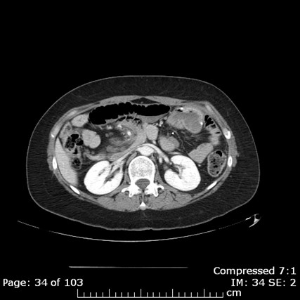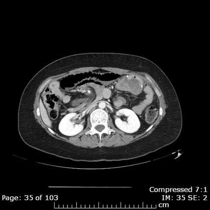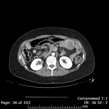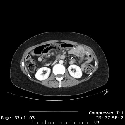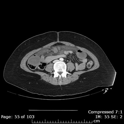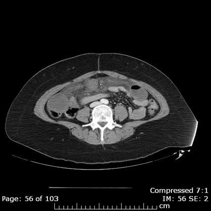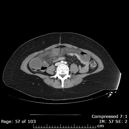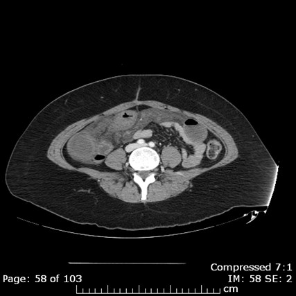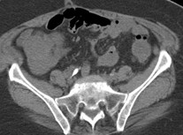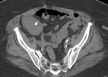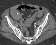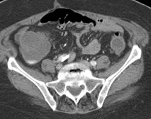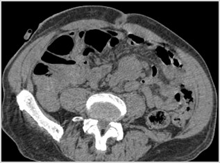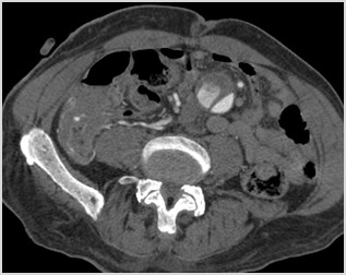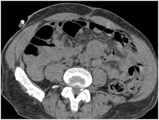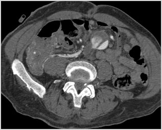
Diagnostic Imaging ofAcute Bowel Pathology
Susan L Summerton, MD

logonew.JPG
http://livingproofmag.com/mag/wp-content/uploads/2011/07/kenny-scharf-phi-living-proof-mag-01.jpg
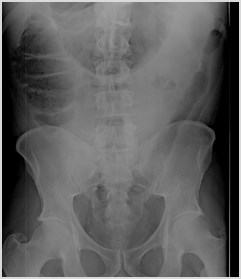
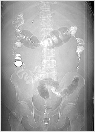
C:\Users\Ryan\Desktop\RADIOLOGY\TEACHING FILES\Bowel\Small bowel obstruction 2\upright plain film.jpg
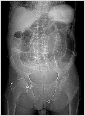
SUPINE
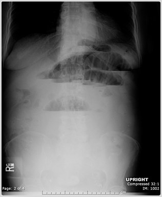
UPRIGHT
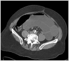
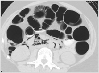
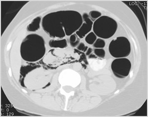
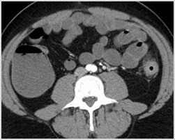
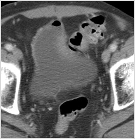
C:\Users\Ryan\Desktop\RADIOLOGY\TEACHING FILES\Bowel\SBO on CT\SBO.ct.01.jpg
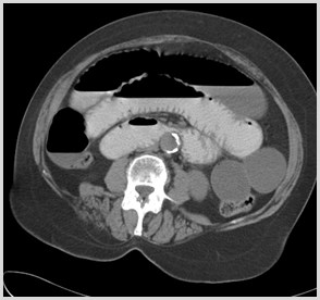
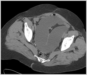
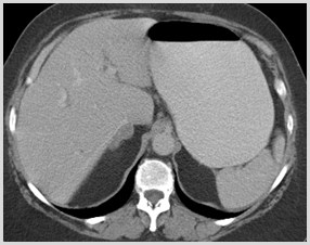
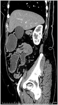
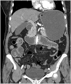
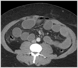
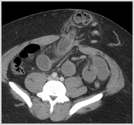
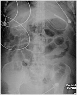
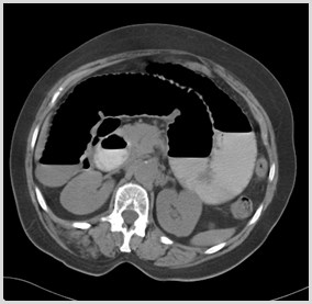
Dilated Loops ofSmall Bowel
Paucity of BowelGas in Colon
Multiple Air-Fluid Levels atDifferent Levels
Small Bowel Obstruction
SBO Imaging
•Plain films
•80-90% vs 30-70% accuracy
•Do not assess site or cause
•CT
•90% accuracy in suspected high grade SBO
•Identifies site and cause
•Most common cause is adhesions
–(with or without history of prior abdominal surgery)
•Identifies complications
•Small-bowel Follow-Through
•Problem solving tool
Contrast
•Oral – Controversial
-Delays diagnosis
-Aspiration risk
-Intravascular volumedepletion/ electrolyteimbalances
+Differentiates partial fromcomplete SBO
+Accelerates surgicaldecision in a completeSBO
•IV
•Always use unlesscontraindications
•Helpful in diagnosingbowel wall ischemia
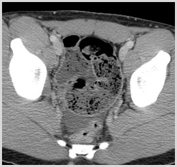
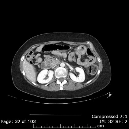
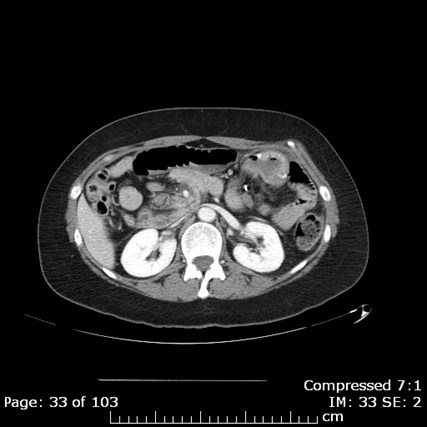
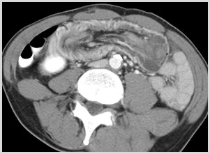
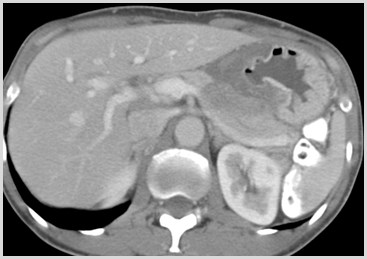
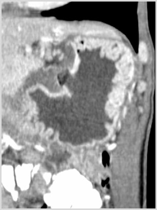
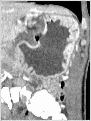
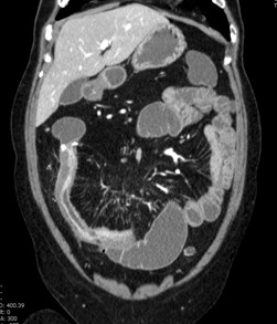
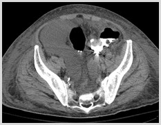
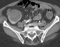
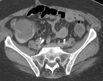
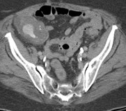
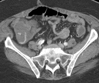
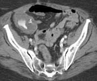
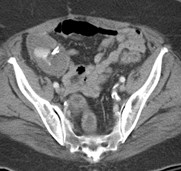
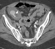
Small Bowel Obstruction:CT Findings
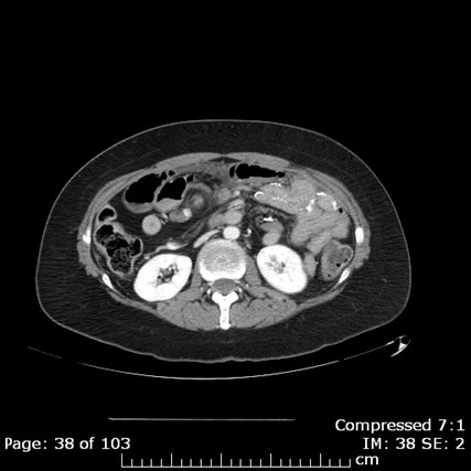
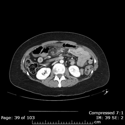
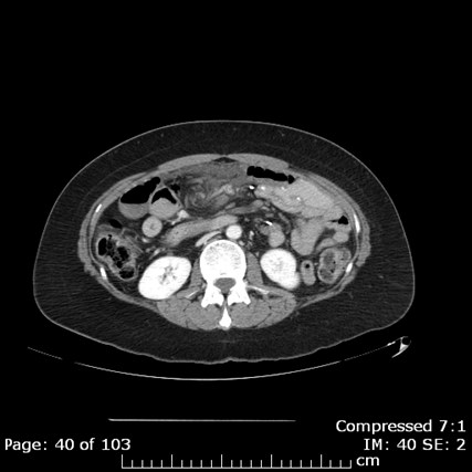
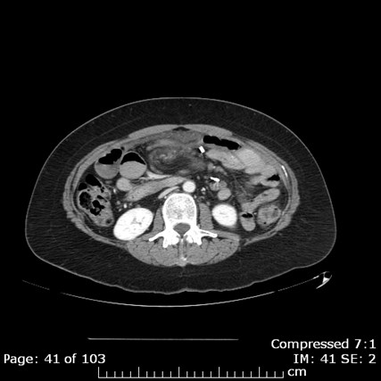
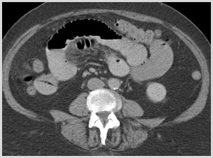
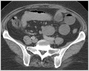
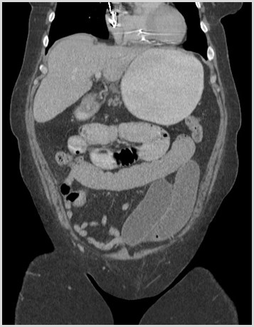
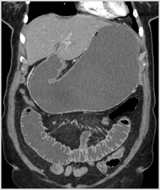
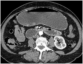
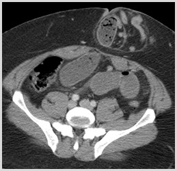
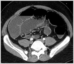
•Dilated loops of small bowel to atransition point
•Multiple air-fluid levels
•Progressive dilution of oral contrast
•Small bowel feces sign
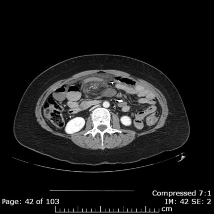
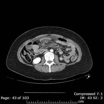
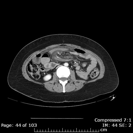
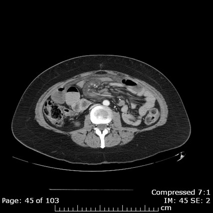
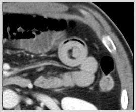
•Whirl Sign
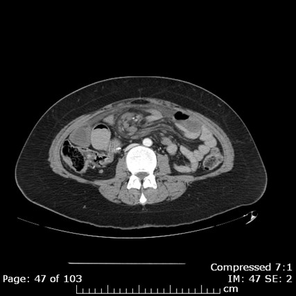
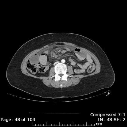
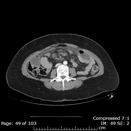
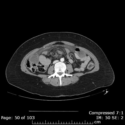
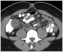
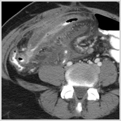
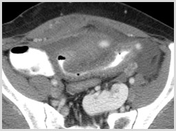
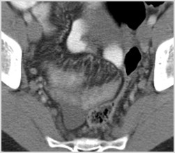
C:\Users\Ryan\Desktop\Desktop Data\Radiology\TEACHING FILES\Bowel\SBO\CT\ser002img00033.jpg
C:\Users\Ryan\Desktop\Desktop Data\Radiology\TEACHING FILES\Bowel\SBO\CT\ser002img00044.jpg
C:\Users\Ryan\Desktop\Desktop Data\Radiology\TEACHING FILES\Bowel\SBO\CT\ser002img00072.jpg
C:\Users\Ryan\Desktop\Desktop Data\Radiology\TEACHING FILES\Bowel\SBO\CT\ser002img00055.jpg
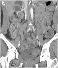
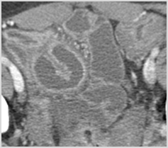
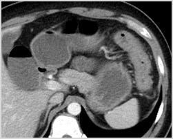
Small Bowel Feces Sign
•Present more frequently inpatients with moderate and highdegrees of SBO
•When present, can be used tolocate the transition point in SBO
Lazarus et al.  Frequency and Relevance ofthe “Small-Bowel Feces” Sign on CT inPatients with Small Bowel Obstruction.AJR 2004; 183: 1361-1366
C:\DOCUME~1\SmithRy\Local Settings\Temp\1.2.840.113619.2.55.3.1510606170.129.1297686851.608.731.bmp
C:\DOCUME~1\SmithRy\Local Settings\Temp\1.2.840.113619.2.55.3.1510605370.730.1295959292.191.751.bmp
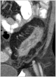
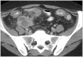
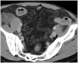
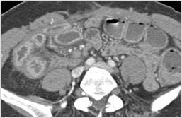
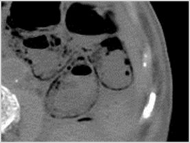
Whirl Sign
•T
w
i
s
t
 o
f
 b
o
w
e
l
 w
r
a
p
p
i
n
g
 a
r
o
u
n
d
 a
 s
i
n
g
l
e
 c
o
n
s
t
r
i
c
t
i
v
e
f
o
c
u
s
 o
f
 m
e
s
e
n
t
e
r
y
•S
i
g
n
 o
f
 c
l
o
s
e
d
 l
o
o
p
 o
b
s
t
r
u
c
t
i
o
n
•I
n
d
i
c
a
t
e
s
 p
o
s
s
i
b
l
e
 c
o
m
p
r
o
m
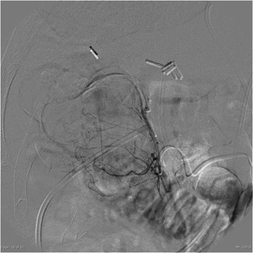
i
s
e
/
c
o
m
p
r
e
s
s
i
o
n
 o
f
v
a
s
c
u
l
a
t
u
r
e
•P
a
t
i
e
n
t
s
 w
i
t
h
 a
 w
h
i
r
l
 s
i
g
n
 o
n
 C
T
 w
e
r
e
 2
5
 t
i
m
e
s
m
o
r
e
 l
i
k
e
l
y
 t
o
 n
e
e
d
 s
u
r
g
e
r
y
 t
h
a
n
 p
a
t
i
e
n
t
s
 w
i
t
h
o
u
t
Duda et al.  Utility of CT whirl sign inguiding management of small bowelobstruction.  AJR 2008; 191: 743-747
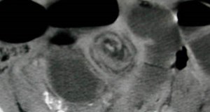
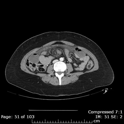
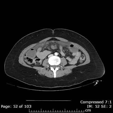
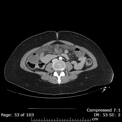
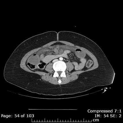
sb volvulus ct kub whirl sign 1a
C:\DOCUME~1\SmithRy\Local Settings\Temp\1.2.840.113619.2.55.3.1477052166.344.1291646822.741.321.bmp
C:\DOCUME~1\SmithRy\Local Settings\Temp\1.2.840.113619.2.55.3.1477052166.344.1291646822.741.331.bmp
C:\DOCUME~1\SmithRy\Local Settings\Temp\1.2.840.113619.2.55.3.1477052166.344.1291646822.741.341.bmp
C:\DOCUME~1\SmithRy\Local Settings\Temp\1.2.840.113619.2.55.3.1477052166.344.1291646822.741.351.bmp
C:\DOCUME~1\SmithRy\Local Settings\Temp\1.2.840.113619.2.55.3.1477052166.344.1291646822.741.361.bmp
C:\DOCUME~1\SmithRy\Local Settings\Temp\1.2.840.113619.2.55.3.1477052166.344.1291646822.741.371.bmp
C:\DOCUME~1\SmithRy\Local Settings\Temp\1.2.840.113619.2.55.3.1477052166.344.1291646822.741.381.bmp
C:\DOCUME~1\SmithRy\Local Settings\Temp\1.2.840.113619.2.55.3.1477052166.344.1291646822.741.391.bmp
C:\DOCUME~1\SmithRy\Local Settings\Temp\1.2.840.113619.2.55.3.1477052166.344.1291646822.741.401.bmp
C:\DOCUME~1\SmithRy\Local Settings\Temp\1.2.840.113619.2.55.3.1477052166.344.1291646822.741.411.bmp
C:\DOCUME~1\SmithRy\Local Settings\Temp\1.2.840.113619.2.55.3.1477052166.344.1291646822.741.421.bmp
C:\DOCUME~1\SmithRy\Local Settings\Temp\1.2.840.113619.2.55.3.1477052166.344.1291646822.741.431.bmp
C:\DOCUME~1\SmithRy\Local Settings\Temp\1.2.840.113619.2.55.3.1477052166.344.1291646822.741.441.bmp
C:\DOCUME~1\SmithRy\Local Settings\Temp\1.2.840.113619.2.55.3.1477052166.344.1291646822.741.451.bmp
C:\DOCUME~1\SmithRy\Local Settings\Temp\1.2.840.113619.2.55.3.1477052166.344.1291646822.741.461.bmp
C:\DOCUME~1\SmithRy\Local Settings\Temp\1.2.840.113619.2.55.3.1477052166.344.1291646822.741.471.bmp
C:\DOCUME~1\SmithRy\Local Settings\Temp\1.2.840.113619.2.55.3.1477052166.344.1291646822.741.481.bmp
C:\DOCUME~1\SmithRy\Local Settings\Temp\1.2.840.113619.2.55.3.1477052166.344.1291646822.741.491.bmp
C:\DOCUME~1\SmithRy\Local Settings\Temp\1.2.840.113619.2.55.3.1477052166.344.1291646822.741.501.bmp
C:\DOCUME~1\SmithRy\Local Settings\Temp\1.2.840.113619.2.55.3.1477052166.344.1291646822.741.511.bmp
C:\DOCUME~1\SmithRy\Local Settings\Temp\1.2.840.113619.2.55.3.1477052166.344.1291646822.741.521.bmp
C:\DOCUME~1\SmithRy\Local Settings\Temp\1.2.840.113619.2.55.3.1477052166.344.1291646822.741.531.bmp
C:\DOCUME~1\SmithRy\Local Settings\Temp\1.2.840.113619.2.55.3.1477052166.344.1291646822.741.541.bmp
C:\DOCUME~1\SmithRy\Local Settings\Temp\1.2.840.113619.2.55.3.1477052166.344.1291646822.741.551.bmp
C:\DOCUME~1\SmithRy\Local Settings\Temp\1.2.840.113619.2.55.3.1477052166.344.1291646822.741.561.bmp
C:\DOCUME~1\SmithRy\Local Settings\Temp\1.2.840.113619.2.55.3.1477052166.344.1291646822.741.571.bmp
C:\DOCUME~1\SmithRy\Local Settings\Temp\1.2.840.113619.2.55.3.1477052166.344.1291646822.741.581.bmp
Whirl Sign
Closed Loop Obstruction
•10% of small bowel obstructions
•Mortality rate: 10-37%
•Complications
•Bowel strangulation
•Vascular compromise
•Ischemia/Infarction
C:\Users\Ryan\Desktop\Desktop Data\Radiology\TEACHING FILES\Bowel\Closed loop obstruction, surgically proven\closed loop obstruction, surgically proven.01.jpg
C:\Users\Ryan\Desktop\Desktop Data\Radiology\TEACHING FILES\Bowel\Closed loop obstruction, surgically proven\closed loop obstruction, surgically proven.04.jpg
C:\Users\Ryan\Desktop\Desktop Data\Radiology\TEACHING FILES\Bowel\Closed loop obstruction, surgically proven\closed loop obstruction, surgically proven.02.jpg
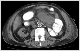
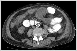
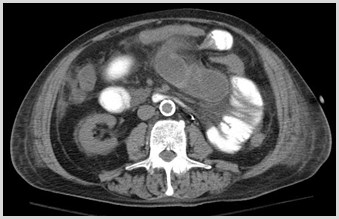
Closed Loop Small BowelObstruction
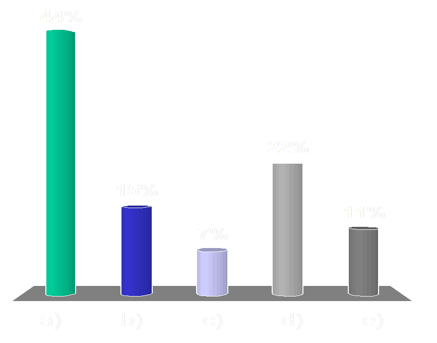
Which of the following causes of a smallbowel obstruction can be diagnosed only onCT and not on x-rays?
Audience Question
a)Adhesions
b)Hernia
c)Mass
d)Gallstone ileus
e)Abscess
Which of the following causes of a smallbowel obstruction can be diagnosed only onCT and not on x-rays?
Audience Question
a)A
d
h
e
s
i
o
n
s
b)H
e
r
n
i
a
c)M
a
s
s
d)G
a
l
l
s
t
o
n
e
 i
l
e
u
s
e)A
b
s
c
e
s
s
Causes of Small BowelObstruction
•Adhesions – only diagnosed on CT
•The adhesive band itself is not visible, butthe finding of abrupt change in caliberwithout other etiology (mass/hernia)implies adhesive band is present
•May be visible on x-rays
•Hernia
•Mass
•Intussusception
•Gallstone ileus
•Abscess/Hematoma
C:\Users\Ryan\Desktop\RADIOLOGY\TEACHING FILES\Bowel\SBO classic findings\CT\ser002img00021.jpg
C:\Users\Ryan\Desktop\RADIOLOGY\TEACHING FILES\Bowel\SBO classic findings\CT\ser002img00044.jpg
C:\Users\Ryan\Desktop\RADIOLOGY\TEACHING FILES\Bowel\SBO classic findings\CT\ser002img00056.jpg
*
Dilated Proximal
Small Bowel
CollapsedColon
Collapsed Distal
Small Bowel
C:\Users\Ryan\Desktop\RADIOLOGY\TEACHING FILES\Bowel\SBO classic findings\CT\ser300img00110.jpg
Proximal Dilated
Small Bowel
Distal Collapsed
Small Bowel
Transition Point
SBO - Adhesion
Intussusception
•Telescoping of one bowel loop into the lumenof an adjacent bowel loop
•Major cause of SBO in children
•Usually no lead point in children
•Adults often have tumor as lead point
•Symptoms
•Crampy abdominal pain
•Bloody stools
•Vomiting
Intussusception: CT Findings
•Alternating bands of high & low density
•Results from intussuscepted mesenteric fatand mucosal-muscular interfaces
•+/- Bowel obstruction
C:\Users\Ryan\Desktop\Desktop Data\Radiology\TEACHING FILES\Bowel\Intussuception\CT axial 1.jpg
C:\Users\Ryan\Desktop\RADIOLOGY\TEACHING FILES\Bowel\SB intussusception\SB intussusception.08.jpg
Do we need to worry about every patientwith intussusception?
•33 adult patients with intussusception
•29/33 small bowel intussusception: only 6 hadneoplastic lead point
•4/33 colon intussusception: all had cancer
•Intussusception due to neoplasm:
•LONGER: 10.8 cm vs 4 cm long
•LARGER: 4 cm vs 3 cm
•More likely to obstruct: 50% vs 4.3%
Warshauer, DM et al. Adult Intussusception Detected at CT or MR Imaging:Clinical-Imaging Correlation. Radiology. 1999;212:853-860
Jejunal mucinousadenocarcinoma
C:\DOCUME~1\SmithRy\Local Settings\Temp\1.2.840.113619.2.5.1260071.5788.1178022768.2111.bmp
C:\DOCUME~1\SmithRy\Local Settings\Temp\1.2.840.113619.2.55.3.1510606170.6001.1178018836.699.3081.bmp
C:\DOCUME~1\SmithRy\Local Settings\Temp\1.2.840.113619.2.5.1260071.5788.1178022768.3121.bmp
C:\DOCUME~1\SmithRy\Local Settings\Temp\1.2.840.113619.2.5.1260071.5788.1178022768.2251.bmp
+
+
+
C:\DOCUME~1\SmithRy\Local Settings\Temp\1.2.840.113619.2.55.3.1510605370.2901.1137153965.963.501.bmp
C:\DOCUME~1\SmithRy\Local Settings\Temp\1.2.840.113619.2.55.3.1510605370.2901.1137153965.963.611.bmp
C:\DOCUME~1\SmithRy\Local Settings\Temp\1.2.840.113619.2.55.3.1510605370.2901.1137153965.963.651.bmp
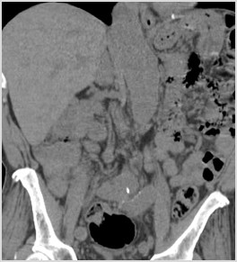
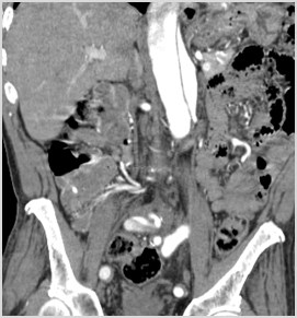
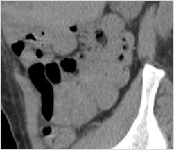
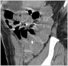
Dilated loops of smallbowel proximal
Small bowel feces sign
SBO
Paraumbilical
Hernia
Decompressed loops
of small bowel distal
SB protrudingthrough abd wall
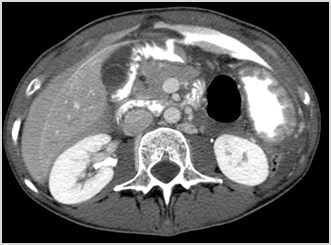
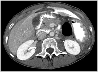
Bowel Perforation
•Small amounts of localized air
•Diverticulitis
•Appendicitis
•Cancer
•Large amounts of air
•Perforated gastric or duodenal ulcer
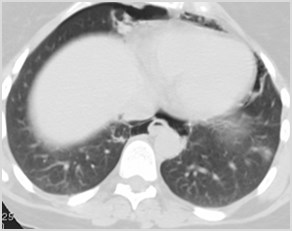
•Retroperitoneal air
•Perforated duodenum
Strong predictors of the site ofbowel perforation
•Focal defect in the bowel wall
•Concentration of extraluminal gas orcontrast adjacent to the bowel wall
•Segmental bowel wall thickening
Hainaux B et al. Accuracy of MDCT in Predicting Site ofGastrointestinal Tract Perforation.  AJR 2006: 1179-1183
C:\Users\Ryan\Desktop\Desktop Data\Radiology\TEACHING FILES\Bowel\Perforated duodenal ulcer\CT axial 1.jpg
C:\Users\Ryan\Desktop\Desktop Data\Radiology\TEACHING FILES\Bowel\Perforated duodenal ulcer\CT axial 2.jpg
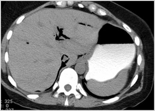
Perforated Duodenal Ulcer
Site of Perforation
Highest Density
Extraluminal Contrast
High Density
Perihepatic Fluid
1
1
1
1
Retroperitoneal air due to duodenal perforation
C:\Users\Ryan\Desktop\Radiology\Penn Cases\GI\gastric ulcer\ct\img001.jpg
C:\Users\Ryan\Desktop\Radiology\Penn Cases\GI\gastric ulcer\ct\img004.jpg
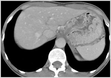
Gastric Ulcer
Gastric Wall Thickening
Gastric Ulcer along the LesserCurvature
C:\Users\Ryan\Desktop\Radiology\Penn Cases\GI\gastric ulcer\ct\img009.jpg
C:\Users\Ryan\Desktop\Radiology\Penn Cases\GI\gastric ulcer\ct\img010.jpg
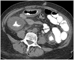
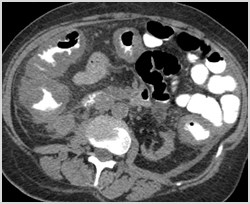
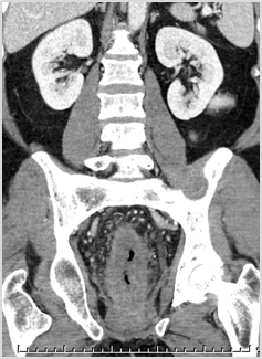
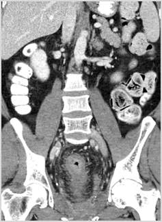
Colitis
•Infectious
•Pseudomembranous colitis
•Inflammatory
•Crohn’s
•Ulcerative colitis
•Ischemic
•Diverticulitis
Colitis
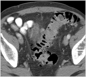
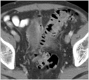
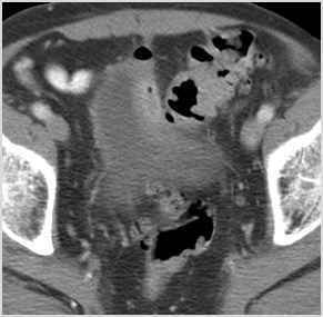
•CT of abdomen and pelvis with oral andintravenous contrast
•Findings are most often nonspecific
•Certain patterns/features on CT canhelp narrow the differential diagnosis
•Clinical and laboratory data
•Colonoscopy and biopsy
C:\DOCUME~1\SmithRy\LOCALS~1\Temp\1.2.392.200036.9125.4.0.185592148.383322880.13856278741.bmp
C:\DOCUME~1\SmithRy\LOCALS~1\Temp\1.2.840.113619.2.55.3.1510605370.220.1273140977.84.551.bmp
C:\DOCUME~1\SmithRy\LOCALS~1\Temp\1.2.840.113619.2.55.3.1510605370.220.1273140977.84.110.bmp
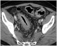
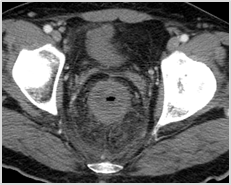
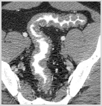
Colonic wall thickening
Radiation Proctitis
C:\DOCUME~1\SmithRy\LOCALS~1\Temp\1.2.840.113619.2.55.3.1510606170.567.1203337543.464.751.bmp
C:\DOCUME~1\SmithRy\LOCALS~1\Temp\1.2.840.113619.2.55.3.1510606170.567.1203337543.464.821.bmp
C:\DOCUME~1\SmithRy\LOCALS~1\Temp\1.2.840.113619.2.5.1260071.9393.1203343152.971.bmp
C:\DOCUME~1\SmithRy\LOCALS~1\Temp\1.2.840.113619.2.5.1260071.9393.1203343152.911.bmp
C:\Users\Ryan\Desktop\Desktop Data\Radiology\TEACHING FILES\Bowel\Pseudomembranous colitis\pseudomembranous colitis.07.jpg
C:\Users\Ryan\Desktop\Desktop Data\Radiology\TEACHING FILES\Bowel\Pseudomembranous colitis\pseudomembranous colitis.08.jpg
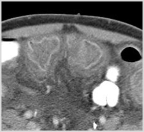
Target Sign
•Alternating bands of density in bowel wall
•Nonspecific/implies acute benign process
•Ischemia/ inflammatory/ infectious
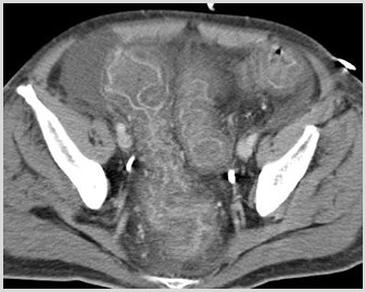
Pseudomembranous Colitis
•Clostridium difficile
•Antibiotic use; Chemo
•Thumbprinting
•Accordion sign
•Ascites
•Often most severe degree ofwall thickening of colitides
C:\DOCUME~1\SmithRy\Local Settings\Temp\1.2.840.113619.2.55.3.1510605370.543.1318767351.502.3101.bmp
C:\Users\Ryan\Desktop\Desktop Data\Radiology\TEACHING FILES\Bowel\Pseudomembranous colitis\pseudomembranous colitis.02.jpg
Crohn’s Disease
•Clinical manifestations
•Abdominal pain and diarrhea
•Weight loss and fever
•Bowel involvement
•Mouth to anus
•Small bowel: terminal ileum most common
•Small and large bowel – 50%
•Large bowel only – 20%
Crohn’s Disease
•Complications
•Bowel obstruction caused by strictures
•Fistulae
•enterocutaneous
•enterovesical
•enterovaginal
•Perforation with sinus tracts
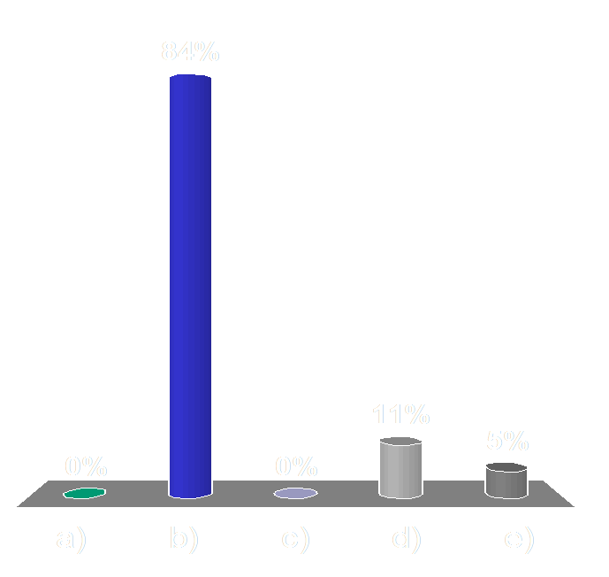
Which of the following studies in evaluating forCrohn’s disease exposes your patient to the leastamount of radiation?
Audience Question
a)CT Enterography
b)MR Enterography
c)Small bowel follow through
d)Barium enema
e)Plain radiographs of theabdomen
Which of the following studies in evaluating forCrohn’s disease exposes your patient to the leastamount of radiation?
Audience Question
a)C
T
 E
n
t
e
r
o
g
r
a
p
h
y
b)M
R
 E
n
t
e
r
o
g
r
a
p
h
y
c)S
m
a
l
l
 b
o
w
e
l
 f
o
l
l
o
w
 t
h
r
o
u
g
h
d)B
a
r
i
u
m
 e
n
e
m
a
e)P
l
a
i
n
 r
a
d
i
o
g
r
a
p
h
s
 o
f
 t
h
e
a
b
d
o
m
e
n
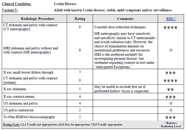
Role of Imaging
•Diagnosis
•Clinical, lab, histology
•Identify location andseverity
•Complications
•Following disease activity
•Monitoring therapy
Routine CT
SBFT
Enteroclysis
CT or MR
Enterography
Crohn’s Disease
Reprinted with permission of the American College of Radiology. No otherrepresentation of this material is authorized without expressed, writtenpermission from the American College of Radiology.
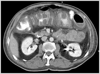
Crohn’s Disease: CT Findings
•Wall thickening with contrast enhancement
•Continuous or segmental  (Skip lesions)
•Narrowed lumen (String sign)
•Increased fat around affected segments ofbowel (Creeping fat)
•Separation of bowel loops (Proud loop)
•Engorged mesenteric vessels (Comb sign)
•Complications: fistulae, abscesses
Crohn’s Disease
C:\DOCUME~1\SmithRy\Local Settings\Temp\1.2.840.113619.2.55.3.1510605370.2929.1120831619.260.451.bmp
C:\DOCUME~1\SmithRy\Local Settings\Temp\1.2.840.113619.2.55.3.1510605370.5796.1174809050.809.481.bmp
C:\DOCUME~1\SmithRy\Local Settings\Temp\1.2.840.113619.2.55.3.1510605370.5796.1174809050.809.611.bmp
String sign
CT Findings
C:\Users\Ryan\Desktop\Desktop Data\Radiology\TEACHING FILES\Bowel\Crohns\_smithry__0804120144\ser002img00068.jpg
C:\DOCUME~1\SmithRy\LOCALS~1\Temp\1.2.840.113704.7.1.1.8244.1351501318.1821.bmp
C:\DOCUME~1\SmithRy\LOCALS~1\Temp\1.2.840.113619.2.55.3.1477050882.431.1355432877.30.641.bmp
C:\DOCUME~1\SmithRy\LOCALS~1\Temp\1.2.840.113704.1.111.5728.1349930678.460931.bmp
Creeping fat
Abscess
Engorged vasa recta
“comb sign”
Crohn’s Disease
C:\Users\Ryan\Desktop\RADIOLOGY\TEACHING FILES\Bowel\chron's enterography\CT\ser003img00128.jpg
C:\Users\Ryan\Desktop\RADIOLOGY\TEACHING FILES\Bowel\chron's enterography\CT\ser004img00092.jpg
C:\Users\Ryan\Desktop\RADIOLOGY\TEACHING FILES\Bowel\chron's enterography\CT\ser003img00076.jpg
Inflamed bowel
Thick walled
Submucosal enhancement
Normal
2T90NCY0_0912081053_0003.JPG
Ulcerative Colitis
C:\DOCUME~1\SmithRy\Local Settings\Temp\1.2.840.113619.2.80.40129220.14269.1316129773.1651.bmp
C:\DOCUME~1\SmithRy\Local Settings\Temp\1.2.392.200036.9125.4.0.152034209.227156224.14530443291.bmp
C:\DOCUME~1\SmithRy\Local Settings\Temp\1.2.840.113619.2.55.3.1510606170.58.1246274385.890.541.bmp
C:\DOCUME~1\SmithRy\Local Settings\Temp\1.2.840.113619.2.55.3.1510606170.58.1246274385.890.281.bmp
Inflammatory Bowel Disease
Ulcerative Colitis
•Left sided or diffuse
•Rectum involved
•Continuous
•Backwash ileitis (dilated)
•No fistulae
•+ Strictures
•+ Abscess
Crohn’s
•Right sided or diffuse
•Rectum often not involved
•Discontinuous
•String sign (narrowed)
•+ Fistulae
•+ Strictures
•+ Abscess
Ischemic Colitis
•Older patients
•Causes
•Arterial occlusion
•Venous occlusion
•Non-occlusive (lower perfusion states)
•Distribution – “Watershed areas”
•distal transverse colon (Griffith’s point)
•distal descending colon (Sudeck’s point)
Ischemic Colitis
•CT angiogram of the abdomen andpelvis utilizing three phases
•Unenhanced
•Arterial
•Venous
•Most specific sign of bowel ischemia
•Non-enhancement of the bowel wall afteradministration of intravenous contrast
Pneumatosis Intestinalis
•High specificity for bowel infarction
•Intraluminal gas dissects throughdisrupted mucosa
•Translocation of bacteria - sepsis
•Gas can progress into
•mesenteric veins
•SMV
•portal veins
C:\Users\Ryan\Desktop\Desktop Data\Radiology\TEACHING FILES\Bowel\ischemic bowel and extensive pneumotosis\ct axial 2.jpg
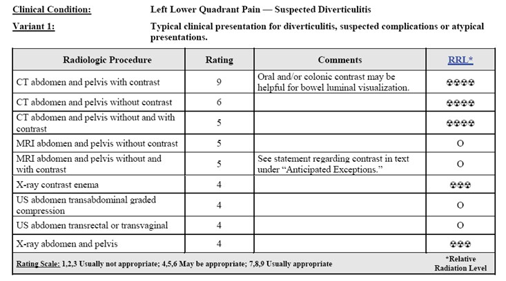
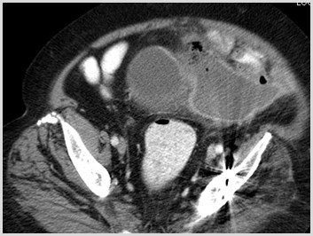
Colonic Diverticulitis
•Outpouchings of colonic mucosa/submucosa that become obstructed
•Most common: descending/sigmoid colon
•Clinical:
•LLQ pain
•elevated WBC
•fever
•+/- bleeding
Reprinted with permission of the American College of Radiology. No otherrepresentation of this material is authorized without expressed, writtenpermission from the American College of Radiology.
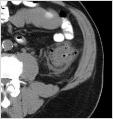
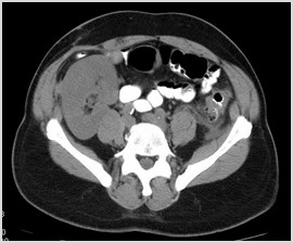
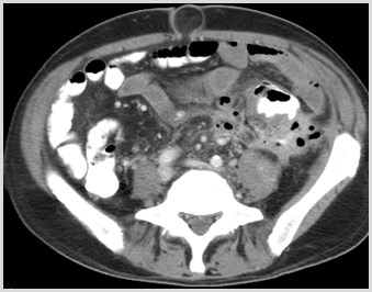
Diverticulitis: CT Findings
•Colonic wall thickening inpresence of diverticula
•Pericolonic fat stranding
•Phlegmon, extraluminal gas,abscess, and dependent freefluid
C:\Users\Ryan\Desktop\Desktop Data\Radiology\TEACHING FILES\Bowel\Diverticulitis\Axial 1.jpg
C:\Users\Ryan\Desktop\Desktop Data\Radiology\TEACHING FILES\Bowel\Diverticulitis\Axial with transplant kidney 3.jpg
Complications of Diverticulitis
•Perforation
•Abscess
•Bowel obstruction
•Fistula formation
•Septic thrombophlebitis
1
1
Complications of Diverticulitis
Perforation and Abscess Formation
Locules of Free Air
Colovesicalfistulae
Acute LowerGastrointestinal Bleeding(LGIB)
Most Common Causes of LGIB
•Diverticular disease (20-55%)
•Angiodysplasia (3-40%)
•Neoplasms (8-26%)
•Colitis (6-22%)
•Benign anorectal lesions (9-10%)
Imaging Options
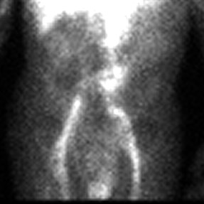
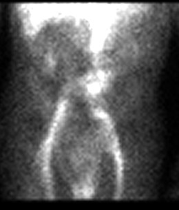
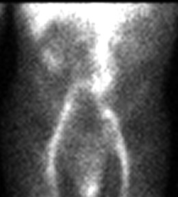
•Nuclear Medicine GI Bleed Study (0.1 mL/min)
•CT Angiography GI Bleed Study (0.3 mL/min)
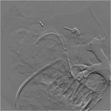
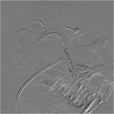
•Conventional Angiography (0.5 mL/min)
•Colonoscopy
•Surgery
CT: Advantages
•A
v
a
i
l
a
b
l
e
 a
n
d
 R
a
p
i
d
l
y
 A
c
q
u
i
r
e
d
•N
o
n
i
n
v
a
s
i
v
e
•P
r
o
v
i
d
e
s
 b
e
t
t
e
r
 l
o
c
a
l
i
z
a
t
i
o
n
•M
a
y
 d
e
f
i
n
e
 t
h
e
 p
a
t
h
o
l
o
g
i
c
 c
a
u
s
e
•E
v
a
l
u
a
t
i
o
n
 o
f
 v
a
s
c
u
l
a
t
u
r
e
•R
e
d
u
c
e
 a
n
g
i
o
 t
i
m
e
 /
 #
 o
f
 r
u
n
s
•R
e
d
u
c
e
 a
n
g
i
o
 c
o
n
t
r
a
s
t
 d
o
s
e
•R
e
d
u
c
e
 a
n
g
i
o
 r
a
d
i
a
t
i
o
n
CT
•Sensitivity: 79-91%
•Specificity: 50-99%
•Diagnostic accuracy: 74-98%
CT: Limitations
•Renal failure
•Prior contrast reaction
•Residual oral contrast in bowel
C:\DOCUME~1\SmithRy\Local Settings\Temp\1.2.840.113619.2.55.3.1510605370.563.1270466920.737.21.bmp
C:\DOCUME~1\SmithRy\Local Settings\Temp\1.2.840.113619.2.55.3.1510605370.563.1270466921.627.491.bmp
C:\DOCUME~1\SmithRy\Local Settings\Temp\1.2.840.113619.2.55.3.1510605370.563.1270466921.627.611.bmp
C:\DOCUME~1\SmithRy\Local Settings\Temp\1.2.840.113619.2.55.3.1510605370.563.1270466921.627.821.bmp
Residual Contrast
Nuclear Medicine: Advantages
•Image over time
•Can use if IV contrast allergy
•Can use with elevated GFR
•Active arterial or venous bleeding ratesas low as 0.1 mL/min
•93% sensitive
•95% specific
Nuclear Medicine: Limitations
•Limited resolution
•Limited availability
•Length of time required to perform study
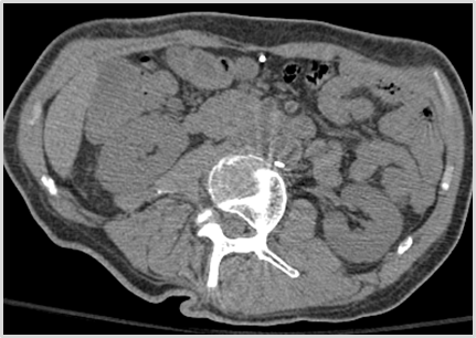
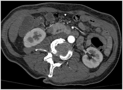
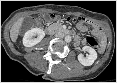
CT of Acute LGIB
•No oral contrast
•Scout – radiologist checks beforeproceeding with rest of study
•Low dose non contrast Abd & Pelvis
•Arterial Phase Abd & Pelvis
•  100-150 cc Optiray 320 at 4 cc/sec
•Venous: Abd & Pelvis -70 sec delay
Diagnosis of Bleeding
•High attenuation material detected within bowellumen at CTA, not present at unenhanced CTperformed immediately prior (Laing et al)
•Linear, jetlike, pooled or swirled or ellipsoid focalcollection of arterial density contrast materialwithin bowel lumen (Tew et al)
1.Tew K, Davies RP, Jadun CK, Kew, J.  MDCT of Acute LowerGastrointestinal Bleeding.  AJR 2004; 182: 427-430.
2.Laing CF, Tobia T, Rosenblum DI et al.  Acute Gastrointestinal Bleeding:Emerging Role of Multidetector CT Angiography and Review of CurrentImaging Techniques.  Radiographics 2007; 27: 1055-1070.
C:\Users\Ryan\Desktop\Desktop Data\Radiology\TEACHING FILES\Bowel\GI Bleed CT\CT\ser006img00052.jpg
C:\Users\Ryan\Desktop\Desktop Data\Radiology\TEACHING FILES\Bowel\GI Bleed CT\CT\ser006img00053.jpg
C:\Users\Ryan\Desktop\Desktop Data\Radiology\TEACHING FILES\Bowel\GI Bleed CT\CT\ser006img00058.jpg
C:\Users\Ryan\Desktop\Desktop Data\Radiology\TEACHING FILES\Bowel\GI Bleed CT\CT\ser002img00052.jpg
C:\Users\Ryan\Desktop\Desktop Data\Radiology\TEACHING FILES\Bowel\GI Bleed CT\CT\ser002img00053.jpg
C:\Users\Ryan\Desktop\Desktop Data\Radiology\TEACHING FILES\Bowel\GI Bleed CT\CT\ser002img00058.jpg
C:\Users\Ryan\Desktop\Desktop Data\Radiology\TEACHING FILES\Bowel\GI Bleed CT\CT\ser002img00059.jpg
C:\Users\Ryan\Desktop\Desktop Data\Radiology\TEACHING FILES\Bowel\GI Bleed CT\CT\ser004img00052.jpg
C:\Users\Ryan\Desktop\Desktop Data\Radiology\TEACHING FILES\Bowel\GI Bleed CT\CT\ser004img00053.jpg
C:\Users\Ryan\Desktop\Desktop Data\Radiology\TEACHING FILES\Bowel\GI Bleed CT\CT\ser004img00058.jpg
C:\Users\Ryan\Desktop\Desktop Data\Radiology\TEACHING FILES\Bowel\GI Bleed CT\CT\ser004img00059.jpg
C:\Users\Ryan\Desktop\Desktop Data\Radiology\TEACHING FILES\Bowel\GI Bleed CT\CT\ser006img00059.jpg
C:\DOCUME~1\SmithRy\Local Settings\Temp\1.2.840.113619.2.55.3.1510605370.611.1265897434.237.1691.bmp
C:\DOCUME~1\SmithRy\Local Settings\Temp\1.2.840.113619.2.55.3.1510605370.611.1265897434.578.1701.bmp
C:\DOCUME~1\SmithRy\Local Settings\Temp\1.2.840.113619.2.55.3.1510605370.611.1265897434.237.1671.bmp
C:\DOCUME~1\SmithRy\Local Settings\Temp\1.2.840.113619.2.55.3.1510605370.611.1265897434.578.1681.bmp
C:\DOCUME~1\SmithRy\Local Settings\Temp\1.2.840.113619.2.80.40129220.3855.1265926944.1531.bmp
C:\DOCUME~1\SmithRy\Local Settings\Temp\1.2.840.113619.2.80.40129220.4456.1265927029.1531.bmp
C:\DOCUME~1\SmithRy\Local Settings\Temp\1.2.840.113619.2.80.40129220.3855.1265926921.771.bmp
C:\DOCUME~1\SmithRy\Local Settings\Temp\1.2.840.113619.2.80.40129220.4456.1265927006.811.bmp
Coronal
SagittalCecum
C:\DOCUME~1\SmithRy\Local Settings\Temp\1.2.840.113845.99.1.3.12.2.1107.5.6.1.10080.31110110021213534651500000002.201.bmp
C:\DOCUME~1\SmithRy\Local Settings\Temp\1.2.840.113845.99.1.3.12.2.1107.5.6.1.10080.31110110021213534651500000002.251.bmp
C:\DOCUME~1\SmithRy\Local Settings\Temp\1.2.840.113845.99.1.3.12.2.1107.5.6.1.10080.31110110021213534651500000002.511.bmp
C:\DOCUME~1\SmithRy\LOCALS~1\Temp\1.2.840.113619.2.55.3.1510605370.961.1359551025.135.1151.bmp
C:\DOCUME~1\SmithRy\LOCALS~1\Temp\1.2.840.113619.2.55.3.1510605370.961.1359551025.499.1201.bmp
C:\DOCUME~1\SmithRy\LOCALS~1\Temp\1.2.840.113619.2.55.3.1510605370.961.1359551026.526.1181.bmp
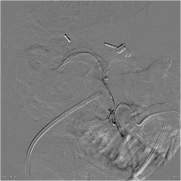
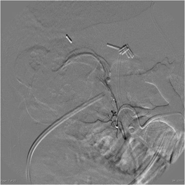
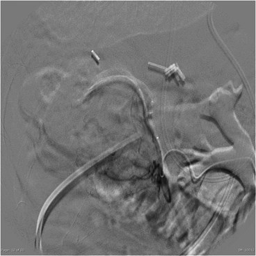
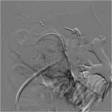
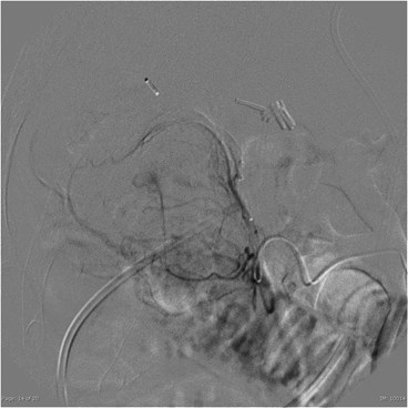
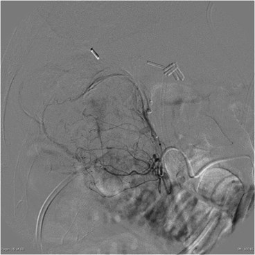
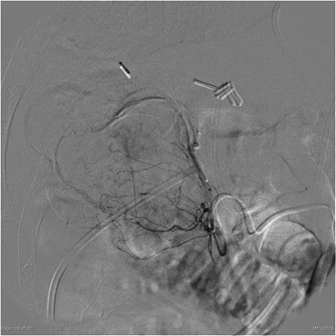
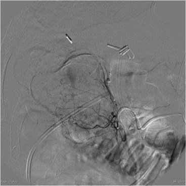
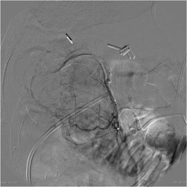
Catheter Angiography
C:\DOCUME~1\SmithRy\LOCALS~1\Temp\1.3.46.670589.7.5.10.80003142025.20130131.135854.16.1.1.10.bmp
C:\DOCUME~1\SmithRy\LOCALS~1\Temp\1.3.46.670589.7.5.10.80003142025.20130131.135854.16.1.4.10.bmp
C:\DOCUME~1\SmithRy\LOCALS~1\Temp\1.3.46.670589.7.5.10.80003142025.20130131.135854.16.1.6.10.bmp
C:\DOCUME~1\SmithRy\LOCALS~1\Temp\1.3.46.670589.7.5.10.80003142025.20130131.135854.16.1.7.10.bmp
C:\DOCUME~1\SmithRy\LOCALS~1\Temp\1.3.46.670589.7.5.10.80003142025.20130131.135854.16.1.12.10.bmp
C:\DOCUME~1\SmithRy\LOCALS~1\Temp\1.3.46.670589.7.5.10.80003142025.20130131.135854.16.1.13.10.bmp
C:\DOCUME~1\SmithRy\LOCALS~1\Temp\1.3.46.670589.7.5.10.80003142025.20130131.135854.16.1.14.10.bmp
C:\DOCUME~1\SmithRy\LOCALS~1\Temp\1.3.46.670589.7.5.10.80003142025.20130131.135854.16.1.15.10.bmp
C:\DOCUME~1\SmithRy\LOCALS~1\Temp\1.3.46.670589.7.5.10.80003142025.20130131.135854.16.1.16.10.bmp
C:\DOCUME~1\SmithRy\LOCALS~1\Temp\1.3.46.670589.7.5.10.80003142025.20130131.135854.16.1.17.10.bmp
C:\DOCUME~1\SmithRy\LOCALS~1\Temp\1.3.46.670589.7.5.10.80003142025.20130131.135854.16.1.18.10.bmp
C:\DOCUME~1\SmithRy\LOCALS~1\Temp\1.3.46.670589.7.5.10.80003142025.20130131.135854.16.1.19.10.bmp
References
1.Small Bowel Obstruction: What to Look For: AC Silva, M Pimenta, LSGuimaraes, RadioGraphics 2009; 29: 423-39.
2.Utility of CT whirl sign in guiding management of small bowelobstruction. Duda et al.  AJR 2008; 191: 743-747
3.Acute Gastrointestinal Bleeding: Emerging Role of Multidetector CTAngiography and Review of Current Imaging Techniques. Laing CF,Tobia T, Rosenblum DI et al. Radiographics 2007; 27: 1055-1070.
4.Detection of active gastrointestinal hemorrhage with CT angiography: a4(1/2)-year retrospective review. Kennedy DW, Laing CJ, Tseng LH,Roseblum DI, Tamarkin SW. J Vasc Interv Radiol 2010; 21(6): 848-855.
5.CT Diagnosis of Acute Mesenteric Ischemia from Various Causes.  AFurukawa, S Kanasaki, et al. American Journal of Roentgenology.2009;192: 408-416.